
Figure 2A to 2D.
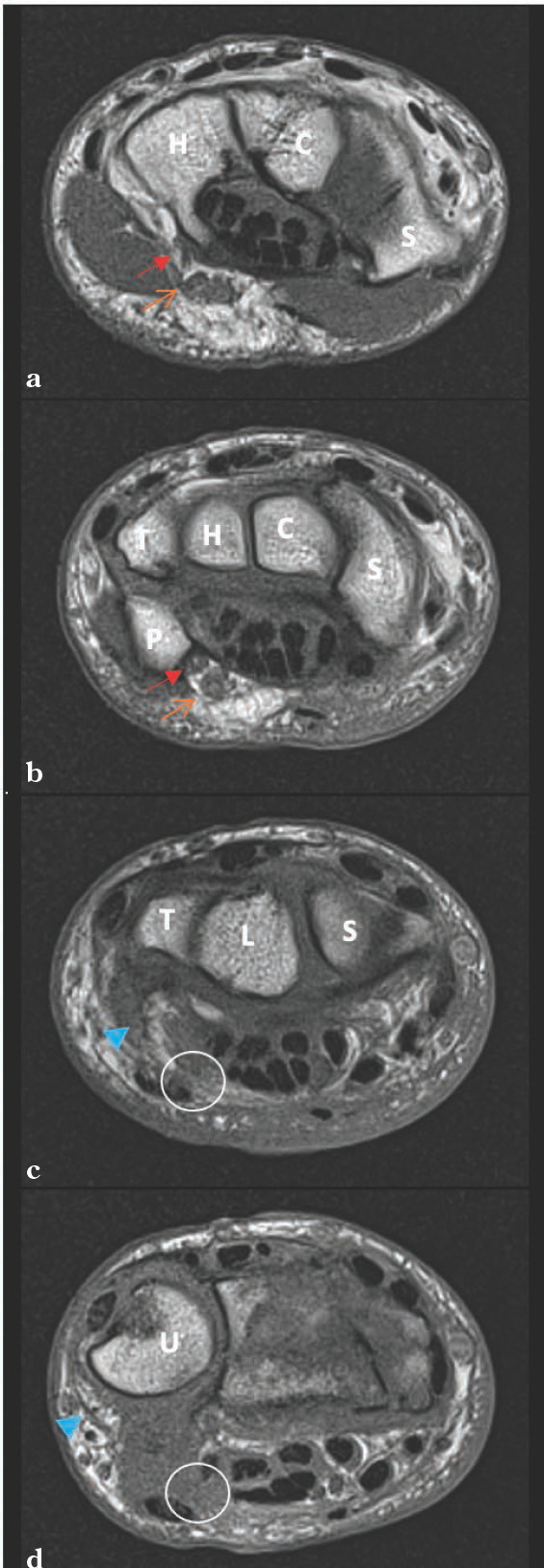
T1 Coronal of the left wrist (2a-2d). Visualized is the deep ulnar nerve becoming confluent with the superficial branch (2a) as the wrist is visualized more proximally (2b). The ulnar neurovascular bundle appears translated ulnarly away from its typical location just radial and deep to the flexor carpi ulnaris. Key: Red solid arrow = deep branch of the ulnar nerve, orange open arrow = superficial branch of the ulnar nerve, solid blue arrowhead = suspected ulnar neurovascular bundle, white circle = normal anatomic location of ulnar nerve, C = capitate, H = hamate, P = pisiform, R = radius, S = scaphoid, T = triquetrum, U = ulna.