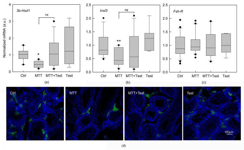
Figure 3.
Effects of mitotane on gene expression in isolated seminiferous tubules and Leydig cells removed from untreated mice (Ctrl) and mice treated with mitotane (MTT), MTT and testosterone (MTT+Test) together, and testosterone (Test). Expression of 3β-Hsd1 (a) and Insl3 (b) in LCs, detected by real-time PCR in LCs within interstitial cells. Expression of Fsh-R (c) mRNA in mouse SCs within the seminiferous tubules detected by real-time PCR. Each sample was normalized to its β-actin content. Results are expressed as arbitrary units (a.u.) and are represented as the mean ± s.e.m. of three independent experiments with total animal numbers of Ctrl = 15, MTT = 18 and MTT + Test = 12, and testosterone = 7. Statistical analysis was performed using ANOVA followed by the Tukey–Kramer test; * p < 0.05 and ** p < 0.01, ns: no Significance. Immunofluorescence analysis (d). Representative images of immunofluorescent 3β-HSD staining in sections of testes from mice treated as described above. Scale bar = 100 μm.

