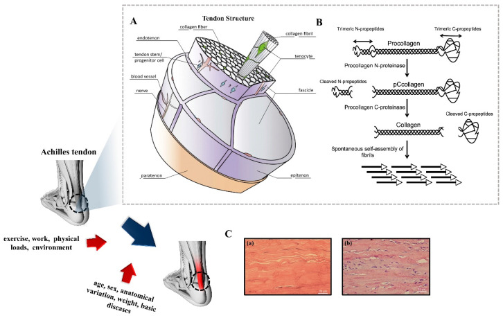
Figure 1.
Hierarchical arrangement of basic tendon structures, pathological changes, and mechanisms of restoration after injury: (A) A schematic drawing of basic tendon structure. Reproduced with permission from Ref. [32]. Copyright 2014, Elsevier B.V., Amsterdam, The Netherlands. (B) Schematic representation of collagen fibril formation by cleavage of procollagen. Reproduced with permission from Ref. [35]. Copyright 2017, International Journal of Experimental Pathology. (C) Histological differences between normal and tendinosis tendon tissue. The normal tendon shows organized collagen fibers and a sparse amount of tenocytes, tightly packed between the collagen bundles (a). In tendinosis (b), the tendon structure becomes disorganized, the tenocytes change morphology and proliferate. Reproduced with permission from Ref. [39]. Copyright 2015, Christensen et al.

