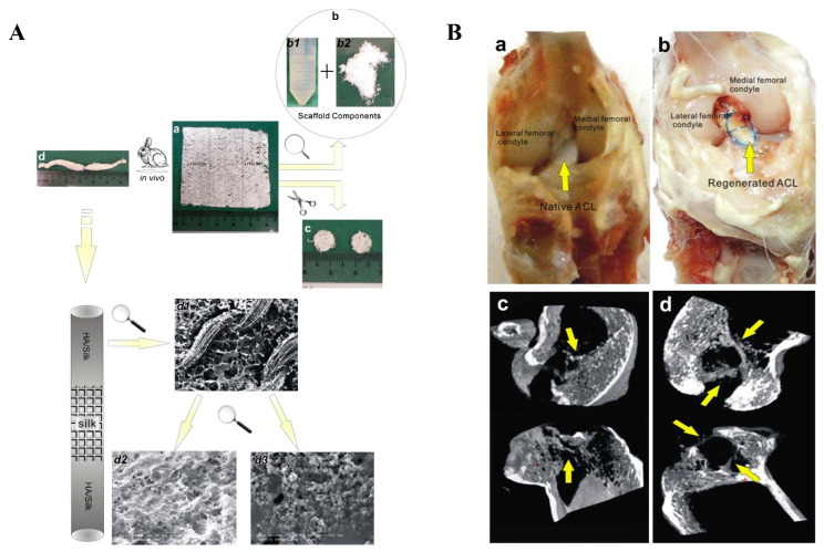
Figure 11.
(A) Diagrammatic sketch of experimental design: gross view of knitted silk mesh with both ends modified by LHA (a); and the components of scaffolds are consisted of silk solution (b1) and LHA (b2); The scaffolds are trimmed in disk shape for in vitro tests (c); the whole knitted silk mesh is rolled up for implantation (d); detailed structure of the rolled-up scaffolds: the scaffold having a highly porous structure (d1), and decoration of CHA and LHA contributes to distinct surface morphology variation (d2,d3). (B) Native ACL (a) and regenerated ACL after 4 months of implantation (b); and m-CT images of implant-bone junction in femoral and tibial bone tunnels after 2 and 4 months implantation (c,d). The yellow arrows point to new bone in the μ-CT images. Reproduced with permission from Ref. [222]. Copyright 2013, Elsevier Ltd.

