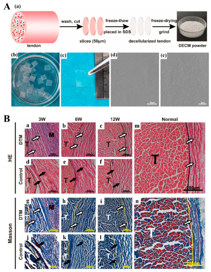
Figure 13.
(A) Preparation and characterization of tendon matrix membranes. Preparation from tendon to DECM powder (a). Representative macroscopic images of wet DTM (b) and dry DTM (c). The surface microstructures of DTM are observed by SEM at magnifications of × 1000 (d) and × 8000 (e). (B) Evaluation of tendon matrix membranes in the repair of a rabbit Achilles tendon. H&E staining (a–f) and Masson staining (g–l) of tissues around tendon after Achilles tendon repair. DTM group, suture with DTM; control group, suture without DTM. H&E staining (m) and Masson staining (n) of normal rabbit tendon without surgery Reproduced with permission from Ref. [244]. Copyright 2021, Elsevier Ltd.

