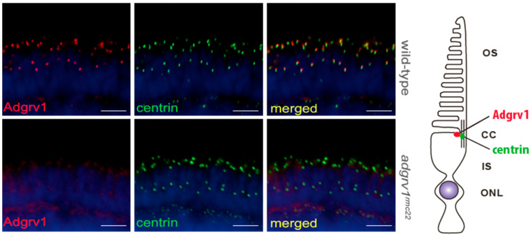
Figure 2.
Localization of Adgrv1 in retinal cryosections of wild-type and adgrv1rmc22 zebrafish. Retinal cryosections of wild-type and adgrv1rmc22 zebrafish larvae (5 dpf) labeled with antibodies directed against Adgrv1 (red) and centrin (green) (as shown by the schematic representation on the right). Nuclei are counterstained with DAPI (blue). In wild-type larvae, Adgrv1 was detected adjacent to the connecting cilium marker centrin, whereas in adgrv1rmc22 zebrafish no Adgrv1 signal could be detected at this location. Scale bar: 10 . OS: outer segment; CC: connecting cilium; IS: inner segment; ONL: outer nuclear layer.

