Figure 4.
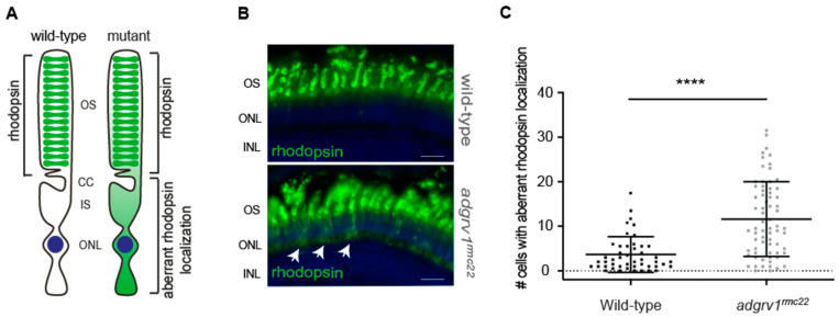
Aberrant localization of rhodopsin in photoreceptor cell bodies in the adgrv1rmc22 zebrafish. (A): Schematic representation of a photoreceptor with rhodopsin localization in the outer segments (OS) versus aberrant rhodopsin localization in the photoreceptor cell body as observed in adgrv1rmc22 mutants. (B): Retinal cryosections of wild-type and adgrv1rmc22 zebrafish larvae (6 dpf) labeled with antibodies directed against rhodopsin (green). Nuclei are counterstained with DAPI (blue). A significantly higher number of photoreceptors with aberrant localization of rhodopsin was observed in adgrv1rmc22 larvae (indicated with the white arrows) than in wild-type larvae. (C): Total number of cells with aberrant rhodopsin localization per retinal section were plotted, with mean SD (n = 29 adgrv1rmc22 mutant larvae and n = 21 wild-type larvae). A two-tailed unpaired Student’s t-test revealed a significant difference between adgrv1rmc22 mutants and wild-types. **** indicates p < 0.0001, scale bar: 10 . CC: connecting cilium; IS: inner segment; ONL: outer nuclear layer; INL: inner nuclear layer.

