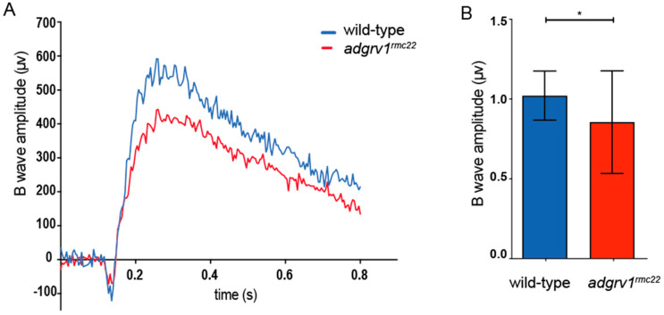
Figure 5.
Electroretinogram recordings reveal impaired retinal function in adgrv1rmc22 juveniles. (A): Representative ERG traces of an adgrv1rmc22 and a wild-type zebrafish. (B): The adgrv1rmc22 zebrafish show a significant decrease in maximum B-wave amplitude when compared to wild-type zebrafish (* p < 0.01, two-tailed unpaired Student’s t-test). ERG traces were recorded on the eyes of juvenile zebrafish (n = 35 adgrv1rmc22 mutants and n = 31 wild-types, 6–8 weeks post fertilization). The average wild-type B-wave amplitude was normalized to 1. Mean B-wave amplitude SD is plotted in the bar graph.

