Figure 3.
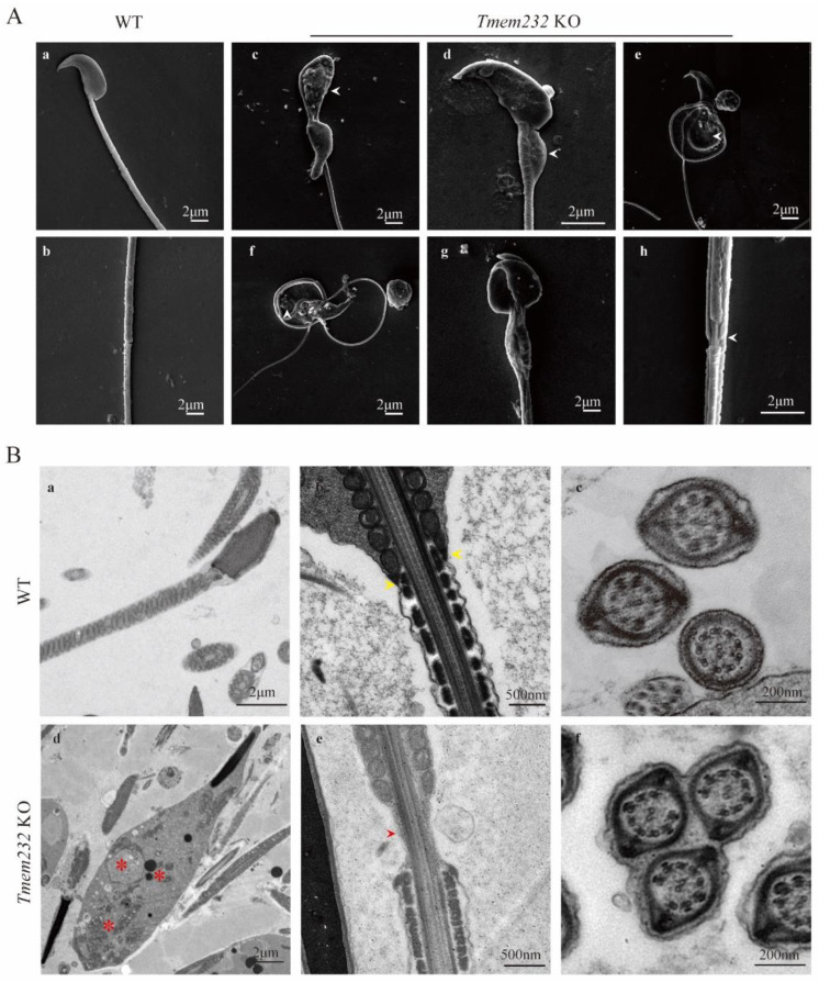
Tmem232 knockout causes failure of the cytoplasm removal. (A) Scanning electron microscopy (SEM) analyses of epididymal spermatozoa of WT and Tmem232 KO mice. WT sperm with normal head and smooth flagella (A), (a). WT sperm with a tight connection between the midpiece and principal piece (A), (b). KO mouse sperm with a large vesicle at the midpiece (A), (c–g), white arrowhead; sperm aggregated into bundles (A), (g,h); sperm with unsheathed flagella at the junction of principal and midpiece piece (A), (h), white arrowhead. (B) Transmission electron microscopy (TEM) analyses of epididymal spermatozoa flagella longitudinal section of WT and Tmem232 KO sperm. WT sperm showed canonical helical mitochondrial sheaths (B), (a), and a tightly connected midpiece and principal piece (B), (b), yellow arrowhead. No sperm bundles were observed in WT mice (B), (c). The abnormal vesicles observed in Tmem232 KO mice displayed several electron-dense materials, disorganized mitochondria, and a single, large vacuole (B), (d), red asterisk. The flagellar longitudinal sections of Tmem232 KO mouse sperm revealed unsheathed flagella at the junction of the principal and midpiece piece (B), (e), red arrow-head. multiple Tmem232 KO mice sperm flagella were wrapped in one cell membrane in cross section (B), (f).

