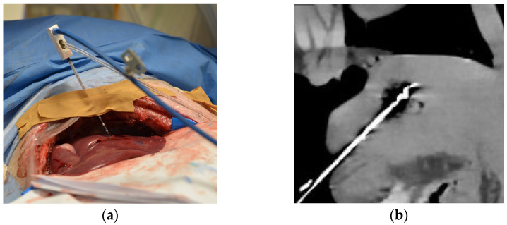
Figure 1.
Experimental setup of computed tomography (CT)-based thermography (CCT): (a) Intraoperative photograph showing the animal under general anesthesia with the liver exposed and the microwave ablation (MWA) probe inserted; (b) CT image showing inserted MWA probe in the liver at the end of heating phase reaching maximum temperature (Tmax) before cooling down. In the same axis with a clear representation of the ablation probe and zone, the images for the rating were set.

