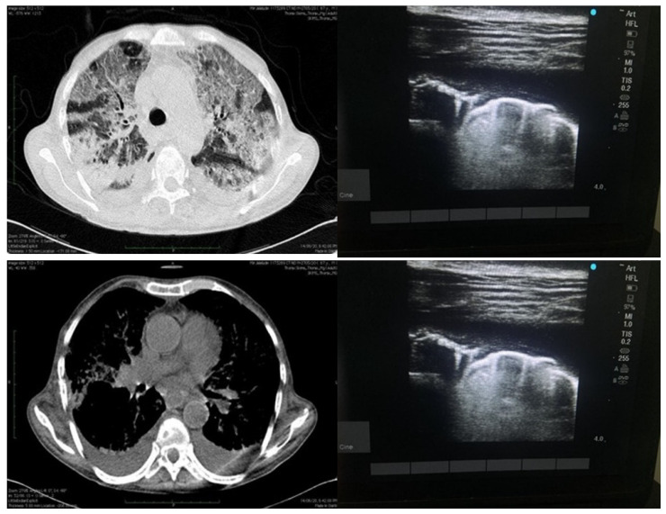
Figure 3.
Depicting findings of LUS on right side (above and below) as effusion with irregular pleural line and isolated B-lines and finally small effusion with isolated B-lines as well as confluent B-lines. Image on the left side depicts same patient with HRCT findings of consolidations as well ground glass opacities and pleural effusion.

