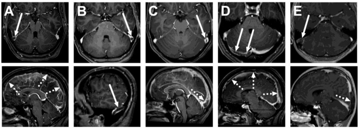
Figure 1.
True positive cases of cerebral venous sinus thrombosis. Axial (top row) and sagittal (bottom row) images of post-gadolinium T1-weighted 3D MRI images of five patients: a 26-year-old female (A), a 33-year-old female (B), a 21-year-old female (C), a 27-year-old female (D), and a 16-year-old male (E). Thrombosis is seen as a hypointense filling defect against the high signal from the gadolinium-based contrast agent in the venous sinuses, usually in the transverse sinuses (arrows) and the superior sagittal sinus (dotted arrows).

