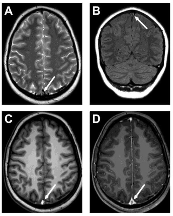
Figure 2.
An example of cerebral venous sinus thrombosis on routine pre-contrast images. Images show a lack of normal flow void on T2-weighted (A) and fluid-attenuated inversion recovery (FLAIR) (B) images, as well as high signal on the pre-contrast T1-weighted image (C), in the superior sagittal sinus (arrows). Post-contrast T1-weighted image is provided for reference (D), showing the non-enhancing thrombus (arrow). Most patients did not show thrombosis on all routine sequences, however. The patient is the same as in Figure 1C.

