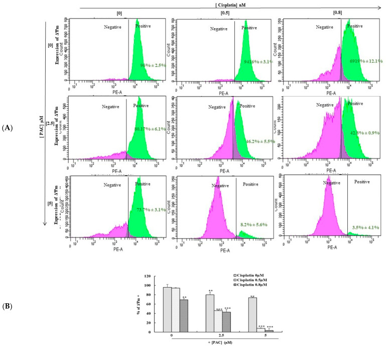
Figure 7.
PAC promotes the effect of cisplatin on the inhibition of mitochondrial membrane potential. (A). (ΔΨm) Expression was measured by DiOC6(3) using flow cytometry. The pink peak represents the % of cells ΔΨm+ cells, and the blue peak is the percentage of ΔΨm− cells (n = 3). ** p < 0.005 *** p < 0.0005 compared to untreated cells. (B) Diagram representing the percentage of positive ΔΨm cells from 3 independent experiments. ** p < 0.05 *** p < 0.0005 compared to untreated cells.

