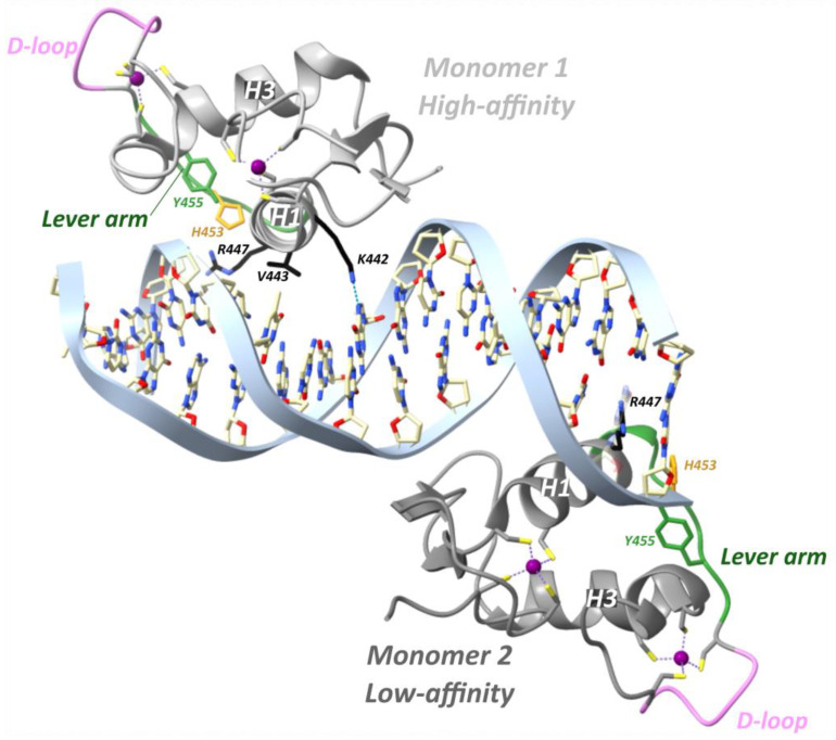
Figure 8.
Crystal structure of the hGR DBD and the TSLP IR-nGRE (PDB ID: 4HN5) [100]. Residues Lys442, Val443 and Arg447 from the first GR monomer and residue Arg447 from the second monomer make specific DNA major groove contacts. His453 is flipped out from the core of the protein fold in both monomers and stabilized by Tyr455 and Arg447. Hydrogen bonds are shown as blue dashed lines; zinc ions are depicted as purple spheres. H1, helix 1; H3, helix 3.

