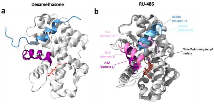
Figure 15.
Structural overview of the dexamethasone-bound and RU-486-bound GR LBD (PDB ID: 1M2Z and 3H52, respectively) [142,151]. (a) Agonist-bound GR LBD as reference with H12 (purple), TIF2 (blue) and dexamethasone (salmon) colored. (b) Overlay of the three domains (domain 1–3) of RU-486-bound GR LBD. In domain 1, RU-486 (salmon) displaces H12 (purple) from the agonist position, enabling the binding of NCOR1 (dark blue). In domain 2, H12 (pink) is observed in an intermediate position between the positions observed in domain 1 and the agonist-bound GR LBD. In domain 3, H12 (light pink) is observed on the other side of the dimethylaminophenyl moiety, occupying the coregulator binding site and thus preventing NCOR1 binding.

