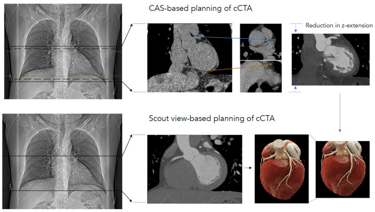
Figure 3.
To obtain a complete depiction of the coronary tree, either calcium-scoring CT-based or scout-view-based planning is feasible. With regard to calcium-scoring CT-based planning (upper row), a calcium-scoring CT scan was planned in anterioposterior (AP) scout view presenting an image 1 cm below the carina and 1 cm below the cardiac apex (solid lines on AP scout view). In the calcium-scoring images, the most cranial part of the coronary tree (blue arrowhead) and the cardiac apex (orange star) are depicted (dotted lines in AP-scout view). CT coronary angiography scan length was set at 1 cm with respect to the cranial and caudal regions of those landmarks (dashed lines on AP-scout view). In scout view-based coronary CT angiography planning (lower row), the scan length was planned based on the scout view, with 1 cm below the carina and 1 cm below the cardiac apex defined as landmarks. The calcium-scoring CT scan itself can be omitted in such an approach.

