
FIG 1.
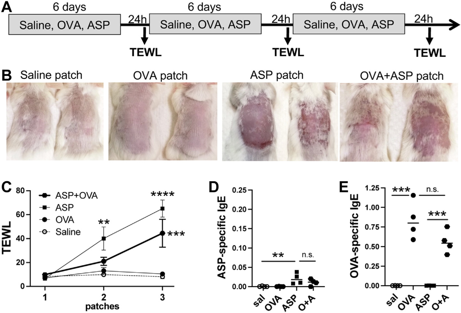
Epicutaneous sensitization to OVA does not require AD-like skin lesions. (A) Protocol: WT BALB/cJ mice were exposed to 3 weekly patches of one of saline, OVA, ASP, or OVA + ASP (1 mg/mL each). (B) Pictures were taken 24 hours after removal of third patch. (C) TEWL was measured 24 hours after patch removal (n = 4 mice per group; significance assessed by 2-way ANOVA). Plasma levels of (D) ASP-specific IgE and (E) OVA-specific IgE (significance assessed by 1-way ANOVA; **P < .01, ***P < .001, ****P < .0001, ns = not significant).