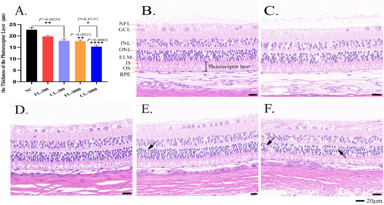
Figure 2.
Effect of LEDs on the morphology of the retinas in albino guinea pigs after 28 days’ exposure. (A) The thickness of the photoreceptor layer (IS and OS) in different groups. Representative retinal H&E stain results show retinal morphology of the NC group (B), FL-300 group (C), CL-300 group (D), FL-3000 group (E), and CL-3000 group (F). NFL, optic nerve fiber layer; GCL, ganglion cell layer; INL, inner nuclear layer; ONL, outer nuclear layer; IS, inner segments of the photoreceptor layer; OS, outer segments of the photoreceptor layer; ELM, external limiting membrane; RPE, retinal pigment epithelium. Black arrows identify the ghost cells. All data are expressed as the mean ± SD (n > 3). **P < 0.01 and ****P < 0.0001 versus the NC group; #P < 0.01 versus the CL-3000 group. Scale bar: 20 µm.

