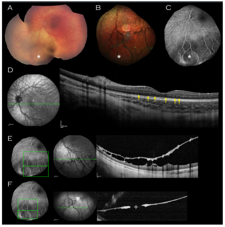
Figure 1.
Left eye imaging of proband. (A) Retcam montage, (B) colour fundus photograph and (C) fluorescein angiogram shows circular area of thinned elevated retina (indicated by asterisk) seen consistently on all imaging modalities. (D) Macular OCT demonstrating unusual thickening of the retina with absence of laminal structure and minute intraretinal hyporeflective cystic lesions (indicated by arrows) and (E,F) OCT through the lesion with approximate mapping of location on fluorescein angiogram image. An associated outer leaf break is demonstrated ((F), cross).

