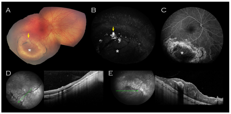
Figure 2.
Right eye imaging of proband. Inferotemporal peripheral island of abnormal retina (indicated by asterisk) on (A) Retcam montage, (B) fundus autofluorescence and (C) fluorescein angiogram. Associated yellow material (arrow (A,B)) was hyperfluorescent on fundus autofluorescence. (D) OCT of the surrounded atrophic retina corresponded with thinned retina and (E) OCT through the centre of the lesion showed thickened retina corresponding to the relatively hypofluorescent island seen on FFA. No schisis was demonstrated on OCT through the lesion.

