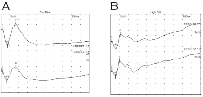
Figure 3.
Electroretinography of proband under general anaesthetic. (A) Dark adapted dim blue scotopic response. (B) Light adapted standard flash (LA 3.0). Waveforms were not electronegative, however, waveforms for right eye (top) indicate relatively reduced b wave amplitudes compared to those for the left eye (bottom).

