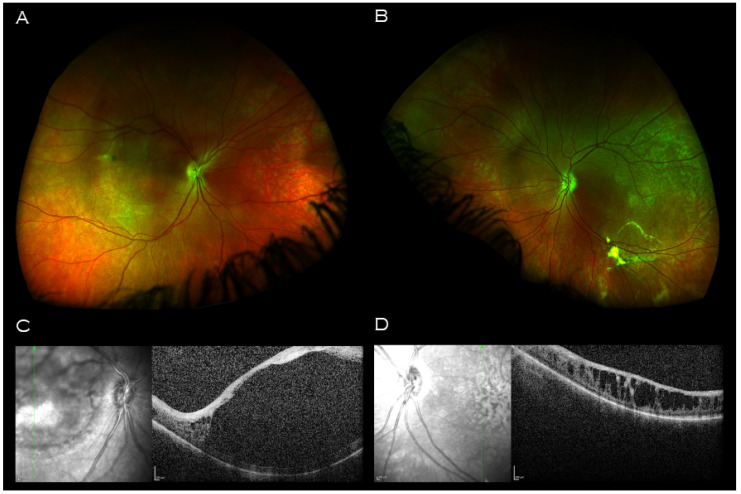
Figure 4.
Imaging of proband’s father. Wide field retinal imaging (A) right eye (B) and left eye shows mild retinal pigment epithelium changes at the maculae and left vitreous veil at left inferior arcade. (C) Macular OCT right eye demonstrating a central hyporeflective intraretinal cavity with adjacent small cystic-like hyporeflective intraretinal lesions peripherally. (D) Macular OCT left eye demonstrating large cystic-like hyporeflective intraretinal lesions throughout, maximal in size and coalescing centrally.

