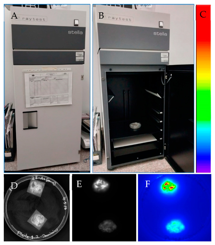
Figure 5.
Luciferase reporter gene system for in vivo detection on banana tissues. (A) Light-tight box linked to a CCD camera used for luciferase activity detection in vivo (Stella 3200, Raytest, Germany). (B) Sample (Petri dish) containing banana embryogenic cells within the light-tight box. (C) Colors indicate luciferase activity. Red indicates high luciferase activity while blue indicates no luciferase activity. (D) Banana embryogenic cells after 3 months of Agrobacterium-mediated transformation using the strain EHA105 harboring the pLVCIBE2 vector (P35S::luc2). Picture was captured in the light-tight box (Stella 3200) under light conditions. The construction of the pLVCIBE2 was as follows: the luc2 was obtained from the pGL4 plasmid (Promega). The gus reporter gene (uidAINT) was digested with the enzymes NcoI (C^CATG_G) and BstEII (G^GTNAC_C) from the pCAMBIA 1301. PCR was performed using the Expand High Fidelity mix (Roche) for the amplification of the luc2 from pGLA4. The primers used were designed with the respective restriction enzyme recognition sequence at the 5′ end (Forward: TAGTACCATGGGGTAAAGCCACCATGGAAGA; reverse: TAGTAGGTCACCCCGCCCCGACTCTAGAATTA). Agrobacterium-mediated transformation was performed according to Santos et al. [38] with some modifications. Briefly, ECS from the banana cultivar ‘Williams’ were developed from male inflorescences. A 33% settled cell volume of 200 μL of ECS (which correspond to approximately 50 mg of fresh banana cells) were co-cultured with 1000 μL of acetosyringone-induced Agrobacterium at OD600nm of 0.4 for 6 h in darkness in a shaker at 25 rpm. Then, banana cells were collected using a 200 μM polyester mesh and subcultured in a ZZ medium for 7 days. Later, cells were subcultured on ZZ medium supplemented with 12.5 mg/L hygromycin and 200 mg/L Timentin®. After one month in a selection medium, luciferase activity was measured. (E) Luciferase activity was detected using the Stella 3200 after adding 20 μL luciferin (500 μM) to the sample, with an acquisition time of 1 min in complete darkness. Image is in greyscale. (F) Acquired image was transformed to pseudo colors.

