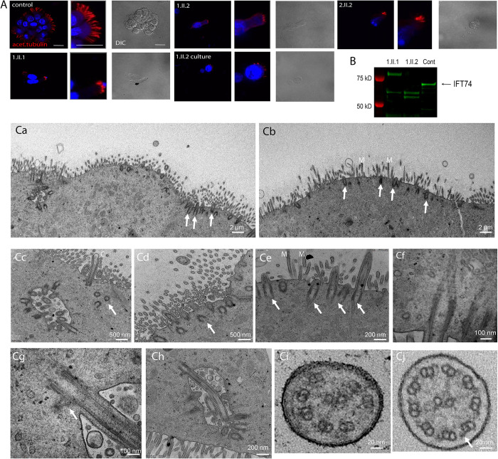
Fig 3. Patients with IFT74 Exon2 Deletion Have Motile Cilia Defects.
(A) Immunofluorescence of respiratory epithelium from a healthy individual (control) and IFT74 exon 2 deletion affected individuals (1.II.1, 1.II.2, 2.II.2). 1.II.2 culture was a biopsy that was cultured for 6 weeks before fixation. Red (acetylated tubulin) marks ciliary axonemes. Blue (DAPI) marks nuclei. Scale bars in the first three images are 20 microns. Corresponding other images in panel are at the same scale. (B) Western blot of nasal biopsy samples. (C) TEM overview images of multiciliated nasal cells (Ca, individual 1.II.2 after cell culture; Cb, 1.II.1 native before cell culture) depicting very short cilia (arrows) mostly not even reaching the length of adjacent microvilli (M). Cc,Cd, close-up image of a ciliated nasal cell in individual 1.II.1 showing shortened cilia (Cc) and several undocked basal bodies below the cell surface (arrows). Ce, short cilia extending from normal docked basal bodes (arrows) in individual 1.II.1. Cf, normal appearing striated ciliary rootlet in 1.II.1. Cg, normal appearing striated basal foot in individual 1.II.1. Ch, example of a longer normally docked cilium in 1.II.1. Ci, ciliary cross sections example of individual 1.II.1 showing microtubule pair misarrangement with missing pairs and no apparent outer dynein arms, indicated with an arrow in a control cross section in Cj.

