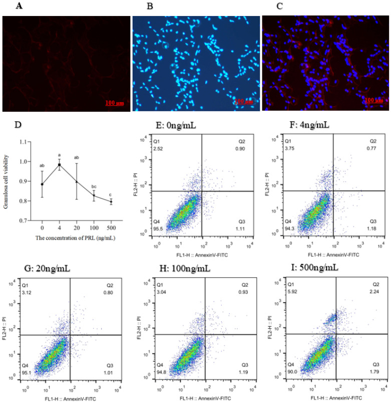
Figure 2.
Identification of ovine ovarian granulosa cells and treatment with different concentrations of PRL on viability (450 nm OD) and apoptosis in cultured ovine ovarian granulosa cells. (A): The red marker denotes the cells expressing FSHR; (B): The blue marker denotes the DAPI-stained nuclei; (C): Merged are a red fluorescently labeled FSHR and a blue fluorescently labeled DAPI overlay; (D): The different lowercase letters indicate significant differences (p < 0.05); (E–I): The X axis represents PI fluorescence; Y axis represents Annexin-V fluorescence. Q1: The dead cells; Q2: The late withered; Q3: The early withered; Q4: The living cells.

