Figure 1.
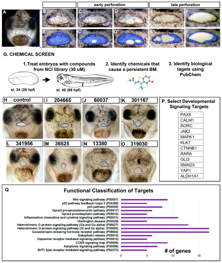
Buccopharyngeal membrane rupture and chemical screen. (A) Frontal view of the face of an embryo at stage 39 prior to the perforation of the buccopharyngeal membrane. (B–F’) magnified images of the mouth at progressive points of buccopharyngeal membrane perforation. In the prime labeled images, the buccopharyngeal membrane is shaded blue. Each image is from a different embryo that represents the most common appearance at each stage (from over 200 embryos examined in 10 biological replicates). (G) Schematic outlining the chemical screen. (H–O) A subset of representative embryos treated with select chemicals causing a persistent buccopharyngeal membrane but with a stomodeum present. The presumptive mouth is outlined in blue dots. The National Service Center Number identifier is shown above each embryo. (P) Protein symbols of select targets of chemicals causing a persistent buccopharyngeal membrane. (Q) The top functional categories identified from the targets of chemicals causing a persistent buccopharyngeal membrane. The GO identifier is in brackets. Abbreviations: # = number, st. = stage.

