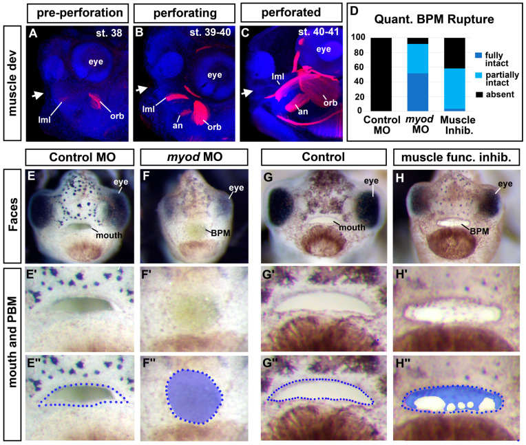
Figure 4.
Cranial muscles are required for buccopharyngeal membrane rupture. (A–C) Lateral views of embryos showing cranial muscles (red) and counterstained with DAPI (blue) (best images from 10 embryos imaged at each stage). White arrows show the location of the developing mouth. (D) Proportion of embryos that had a fully intact, partially intact, or absent buccopharyngeal membrane after myoD knockdown or treatment with a muscle inhibitor (BTS) compared to controls (n = 60, 3 biological replicates for each treatment). (E–H’’) Frontal views of representative embryos injected with myoD morpholinos or treated with an inhibitor of muscle function (BTS). Prime-labeled images show magnified images of the mouth, and the double-prime-labeled images show the buccopharyngeal membrane shaded in blue. Abbreviations: levator mandibulae longus = lml, orbithyoidus = orb, angularis = an, BPM = buccopharyngeal membrane.

