Figure 5.
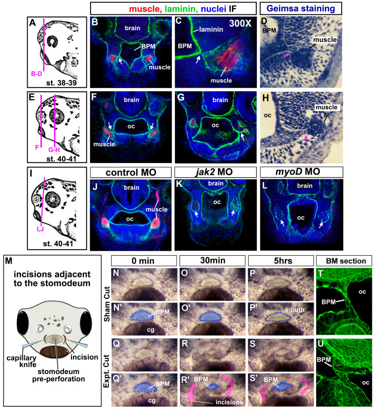
(A–L) Muscle and oral cavity connections. (A,E,I) Schematics of the lateral views of embryos showing locations of the corresponding sections. (B,C,F,G,J–L) Transverse sections labeled with antibodies to detect muscle-specific protein (12/101 in red), laminin (green), and counterstained with DAPI (blue). Images were chosen from representative images taken from 20 different embryos in 2 biological replicates. White arrows indicate laminin that bridges the oral cavity and muscle compartments. (D,H) Plastic section stained with Giemsa showing representative images of the association between muscle and oral cavities (based on sections of 20 embryos in 2 biological replicates). Pink arrows indicate the connections between the muscle and the oral cavity. (M) Schematic of a frontal view showing the location of the surgical incisions. (N–S’) Shows the embryonic mouth and buccopharyngeal membrane in representative embryos before (0 min) and after sham or incisions (at 30 min and 5 h). Prime images show the buccopharyngeal membrane shaded in blue and the mouth outlined in blue dots. Representative images chosen from 50 embryos performed over 5 biological replicates. In R and S, the incised tissue is colored pink. (T,U) representative embryos 30 min after surgeries (or sham) sectioned and labeled with phalloidin to show the cellular arrangements in the buccopharyngeal membrane. Shows representative images taken from a total of 10 embryos in 2 biological replicates, Abbreviations: oc = oral cavity, BPM = buccopharyngeal membrane, cg = cement gland.

