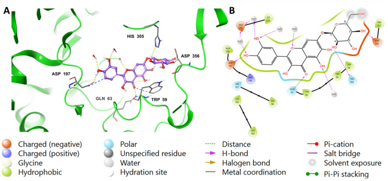
Figure 4.
Binding mode of (2) in the active site of pancreatic alpha-amylase (PDB ID: 4GQR). Compound 2 is shown as purple sticks, while hydrogen bonds and ionic bonds are represented by yellow and blue dotted lines, respectively. (A) A 3D representation of pancreatic alpha-amylase complexed with (2), and (B) a 2D depiction.

