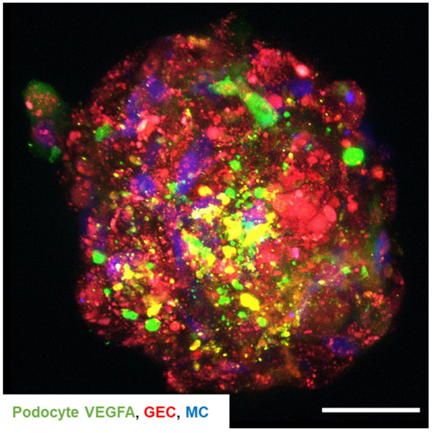
Figure 9.
Podocyte-derived VEGFA is transported to glomerular endothelial cells in 3D glomerular co-cultures. Confocal microscopy of 3D glomerular co-culture displays green fluorescent podocyte-derived VEGFA, mesangial cells labeled in blue with eBioscience™ Cell Proliferation Dye eFluor™ 450 and red fluorescent tdTomato-Farnesyl glomerular endothelial cell reporter cells. Podocytes were electroporated with a plasmid carrying human VEGFA sequence coupled with the GFP sequence, resulting in the expression of green, fluorescent VEGFA prior to co-culture. Scale bar 50 µm.

