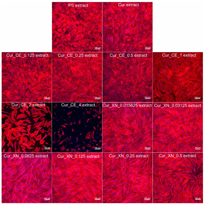
Figure 9.
Morphology of human skin fibroblasts after 4 days of incubation with selected extracts of curdlan-based biomaterials enriched with CE and XN. Extracts from polystyrene (PS) and non-modified curdlan-based biomaterial (Cur) served as controls. Cell nuclei were stained with Hoechst 33342 dye (shown as blue fluorescence) and actin filaments of the cytoskeleton were stained with AlexaFluorTM 635 Phalloidin dye (shown as red fluorescence). Images were obtained using a confocal laser scanning microscope (Olympus Fluoview equipped with FV1000, Shinjuku, Japan); magnification × 100, scale bar 150 μm.

