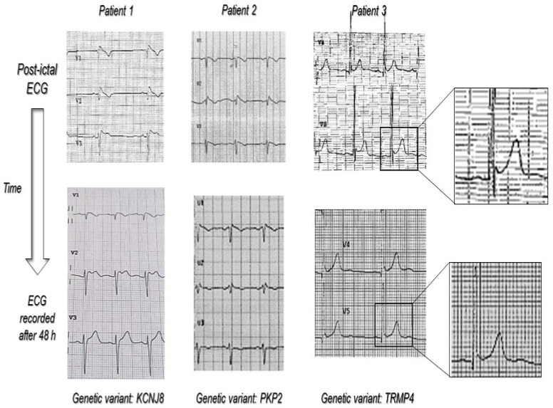
Figure 2.
The figure shows, in detail, the ECG behavior from the post-ictal to the basal (>48 h after the seizure) time in patients with ECG modification diagnostic for myocardial channelopathy (BrS and ERS) and the respective pathogenic gene variant found using NGS analysis. The magnification in patient 3 shows how in the post-ictal ECG, the ERP was more evident. Immediately after seizure, there was a “notching” in the terminal portion of QRS and a 1 mm ST segment elevation, which disappeared in basal ECG.

