
Figure 5.
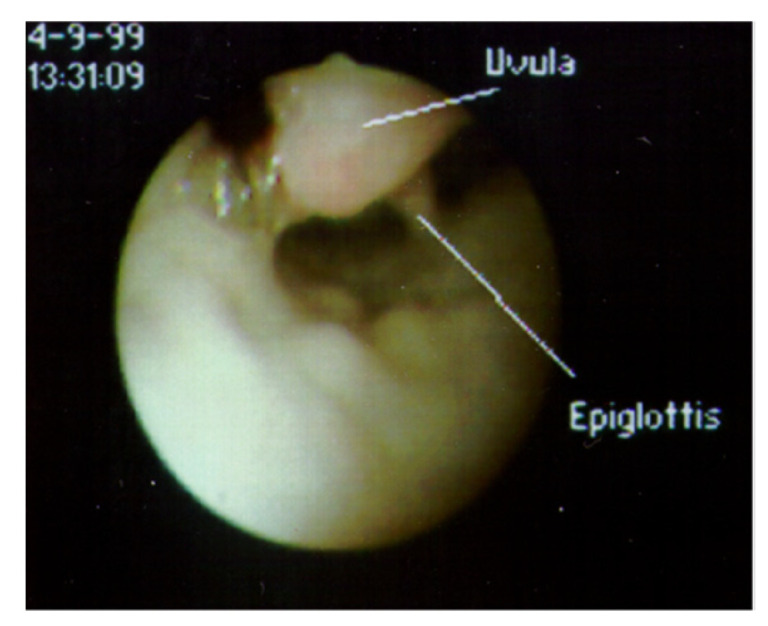
View of supraglottic area through a flexible fiberoptic bronchoscope inserted through the nose, with the patient supine. The uvula, which cannot normally be observed when the epiglottis is visualized, is lying in contact with the epiglottis.