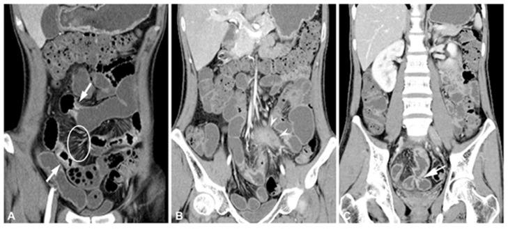
Figure 2.
Diagnosis of inflammatory Crohn’s disease with extraenteric complications by computed tomography enterography. (A) Multifocal segmental stricture showing wall thickening, mural hyperenhancement, and stratification shown by arrows and engorged vasa recta, comb sign highlighted by a circle. (B) Mesenteric abscess is shown by arrowheads. (C) Adjacent to the enteroenteric fistula shown by the arrow. Reprinted with permission of [57].

