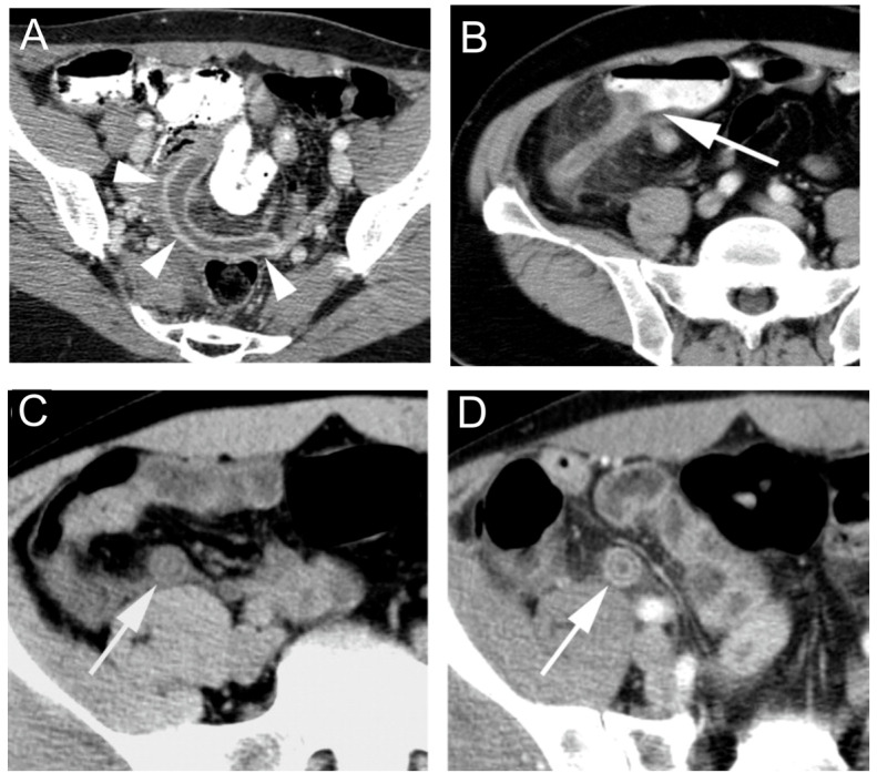
Figure 4.
Computed tomography findings of appendicitis. (A) Axial CT image showing an enlarged, fluid-filled appendix (arrowheads). (B) Axial CT image showing edema of the cecal tip with oral contrast pointing (arrow) towards the base of the inflamed appendix. Axial CT images in the same patient (C) before and (D) after intravenous contrast show a thickened appendix with submucosal edema. Reprinted with permission of [138].

