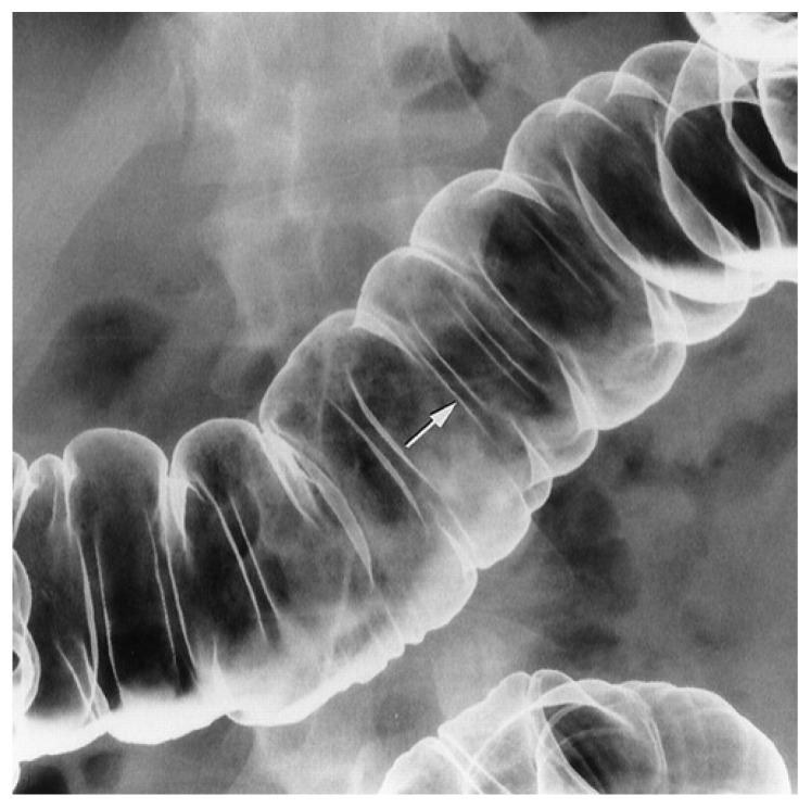
Figure 6.
Spot radiograph of the middle of the transverse colon obtained from a patient near-erect position. The interhaustral folds are straight; a representative fold is identified with an arrow. The haustral sacculations are distended, but not overdistended and flattened. Reprinted with permission of [135].

