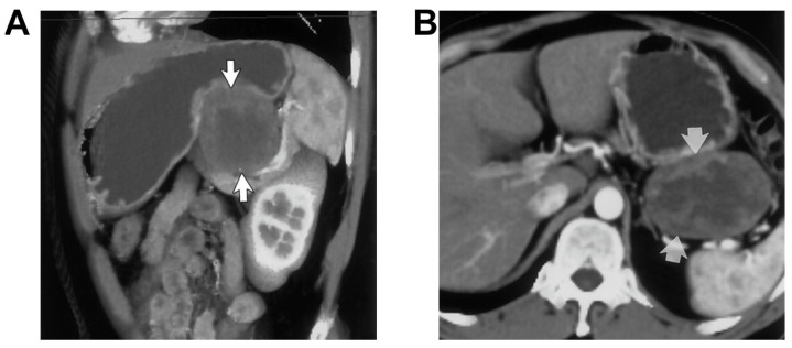
Figure 7.
Sagittal (A) and axial (B) oblique contrast-enhanced 3D volume-rendered CT scans revealed a round exophytic mass in the stomach, 5-cm exophytic mass (arrows) that arises from the stomach, which proved to be a benign GIST during surgery. Reprinted with the permission of [183].

