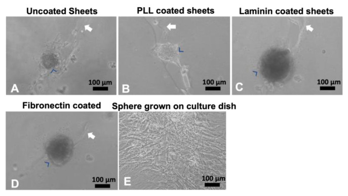
Figure 3.
Human MSCs grown on coated OPF+ sheets containing ridges. Live culture images taken at 10x bright-field. Human MSCs adhered in cell clusters (blue arrowheads) when grown for 3 days on uncoated (A), poly-L-lysine-coated (B), laminin-coated (C), or fibronectin-coated (D) OPF+ ridged sheets (white arrow indicates the ridge). If these spheres were picked and grown in a culture dish, they spread into their stereotypical shape (E).

