Figure 7.
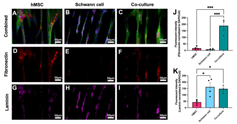
Attachment, polarization, and ECM deposit by hMSCs and rat Schwann cells on patterned 30 × 30 TiSAMP OPF. hMSCs (A) and Schwann cells (B) were cultured on the chemically patterned OPF individually and in co-culture (50:50) on the chemically patterned OPF for 7 days. Using immunocytochemistry, the scaffolds were stained for DAPI (blue), phalloidin (green; (A–C)), fibronectin (red, (D–F)) and laminin (pink, (G–I)) to compare ECM production. Fibronectin (J) and laminin (K) intensities were quantified using ImageJ via normalizing fibronectin production to DAPI/cell number (n = 4 scaffolds with 4 areas per sheet). Statistical analysis of fibronectin staining was run using a one-way ANOVA with multiple comparisons using Tukey’s post hoc test. The degrees of significance are indicated as follows: p < 0.05 (*) and p < 0.001 (***). Error bars represent standard error, individual data points are displayed for each bar.

