Figure 10.
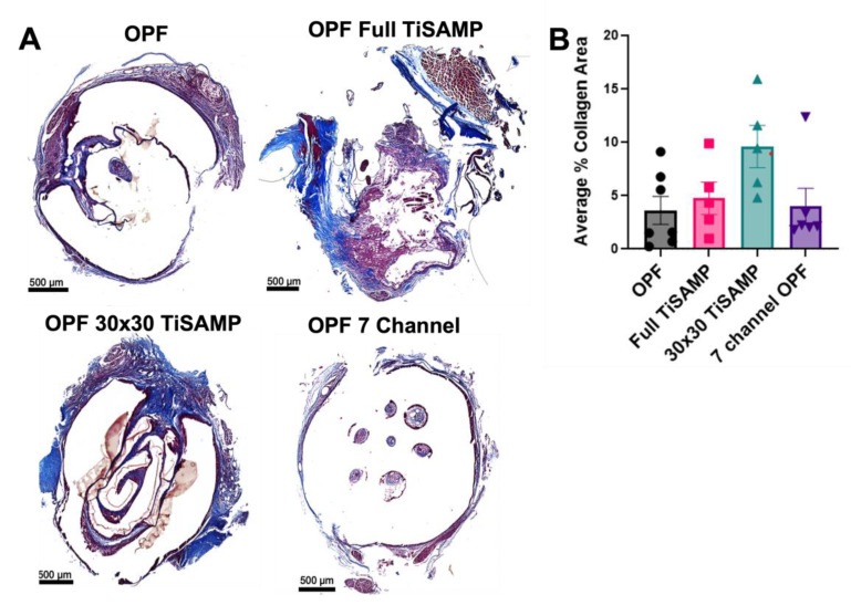
Fibrotic scarring following implantation of OPF scaffolds. (A) Implanted untreated OPF, Full TiSAMP, 30 × 30 TiSAMP, and seven-channel scaffolds were sectioned and stained at quarter lengths (¼, ½, and ¾ of the scaffold; center or ½ segment shown here) with Trichrome histological stain. The collagen component is indicated by blue staining, whereas the cytoplasm is pink. The dark blue stained parts represents areas containing excessive collagen deposits characteristic of fibrotic scarring, with light purple being the overlap of regular collagen deposits within the tissue. A machine learning algorithm was used to analyze the average percent area of collagen scarring at quarter lengths of the scaffold and then averaged (B). There was no significant difference between the conditions. One-way ANOVA with Tukey’s multiple comparisons were used. Error bars represent standard error, individual data points are displayed for each bar.

