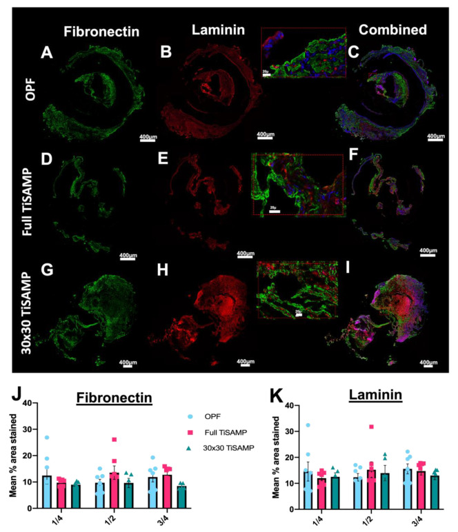
Figure 11.
Fibronectin and laminin deposits by infiltrating cells following implantation of rolled OPF scaffolds. Quarter lengths (¼, ½, and ¾ of the scaffold; center or ½ segment shown here) of untreated rolled OPF scaffold (A–C), full TiSAMP OPF (D–F), and 30 × 30 TiSAMP (G–I) were stained for fibronectin and laminin. Magnified segment of the region of interest in the combined images (C,F,I) are shown in the red box. There was no significant difference in the mean percent area stained through the whole length of the scaffold (One-way ANOVA with Tukey’s multiple comparisons) of either fibronectin (J) or laminin (K). Error bars represent standard error, individual data points are displayed for each bar.

