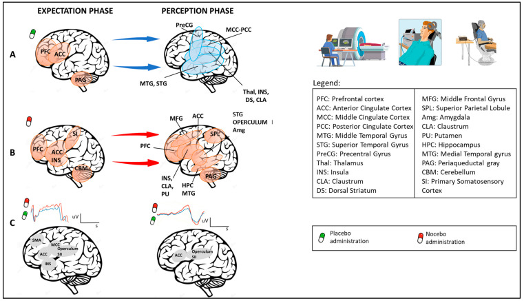
Figure 2.
Neuroimaging studies: temporal aspects (expectation and perception phases) related to brain area activity after placebo or nocebo administration. As reported by different neuroimaging studies, expectations of pain relief, triggered by placebos, activate brain areas such as PFC, ACC, and PAG (P1); in the perception phase, deactivation has been found in different brain regions, including MCC, PCC, MTG, STG, PreCG, Thal, INS, CLA, and DS (A). On the contrary, expectations of pain worsening, triggered by nocebos, enhance activity in brain regions that include PFC, ACC, INS, SI, and CBM; in the perception phase, increased activity in PFC, ACC, MFG, INS, CLA, PU, HPC, MTG, SPL, STG, OPERCULUM, and INS has been found (B). Electroencephalographic (EEG) studies report that placebos and nocebos change EEG brain activity. In particular, the expectation of receiving no painful or painful stimuli respectively decreases (green line) or increases (red line) the amplitude of the contingent negative variation (CNV). Considering the “perception phase”, placebo treatments produce a decrease (blue line) in laser-evoked potential (LEP), an EEG wave that represents an early measure of nociceptive processes (C).

