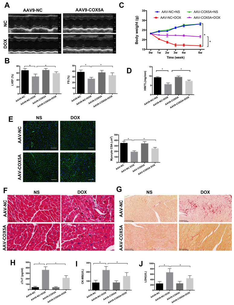
Figure 3.
COX5A protects against DOX-induced cardiomyopathy in mice. (A) Representative M-mode echocardiograms for each group 6 weeks after DOX injection. (B) Echocardiographic quantification of LVEF and FS. (C) Body weight alterations with DOX treatment for different durations. (D) Statistical analysis of HW/TL. (E) Representative WGA-Alexa Fluor 488 conjugate stained heart sections and analysis of cardiomyocyte cross-sectional areas (CSA). Scale bar = 100 μm. (F) Representative histopathological findings of heart sections stained with H&E. Black arrows indicate extensive cytoplasmic vacuolization. Scale bar = 50 μm (G) Representative histopathological manifestations of heart sections stained with Sirius Red. Scale bar = 100 μm. (H) Analysis of serum level of cTnT by Elisa. (I,J) Biochemical determination of CK-MB and LDH serum levels. Data are expressed as mean ± SEM. * p < 0.05.

