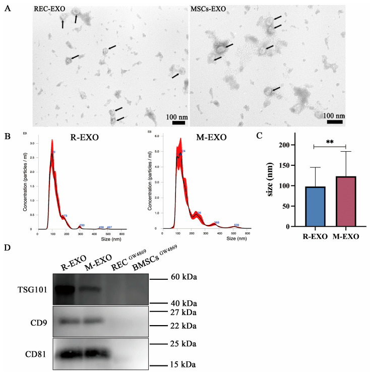
Figure 5.
Identification of exosomes isolated from RECs and BMSCs. (A) Representative TEM images showing round or oval shapes and varied sizes of R-EXO and M-EXO with bilayer membrane structures. Black arrows: exosomes. (B) NanoSight was used to assess the particle size distribution of R-EXO and M-EXO. Error bars indicate ±1 standard error of the mean. (C) Quantitative analysis of particle diameter size (n = 3). Data represent the mean ± standard deviation of three independent experiments. ** p < 0.01. (D) Western blotting for detection of the exosomal markers (TSG101, CD9, and CD81) in R-EXO and M-EXO. R-EXO: REC-derived exosomes. M-EXO: BMSC-derived exosomes. GW4869 is an inhibitor of exosome biogenesis/release.

