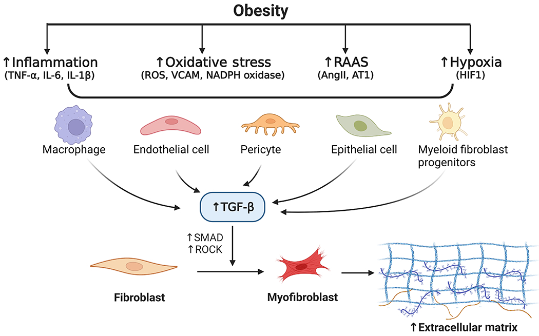
Figure 2. Potential mechanisms of pathological ECM remodelling in obesity.

The molecular and pathophysiological mechanisms that underly maladaptive ECM remodelling in obesity are depicted. Obesity-related inflammation, oxidative stress, RAAS, and hypoxia have been shown to stimulate and activate ECM-producing fibroblast cells, promoting matrix synthesis. Obesity induces inflammation with elevated levels of TNF-α, IL-1, and IL-6. Oxidative stress in obesity is manifested by increases in the ROS levels. Activation of RAAS also leads to increases in ROS via inducing the expression of NADPH oxidase. The expression of vascular cell adhesive molecule (VCAM) is upregulated by ROS and HIF1, which is induced in response to hypoxia. These signals could increase TGF-β expression and initiate TGF-β mediated pro-fibrotic response in various cells including macrophages and endothelial cells. TGF-β has been identified as an important regulator of maladaptive ECM remodelling, promoting excess deposition of ECM components possibly through activation of signalling molecules like SMAD and Rho-associated protein kinase (ROCK).
