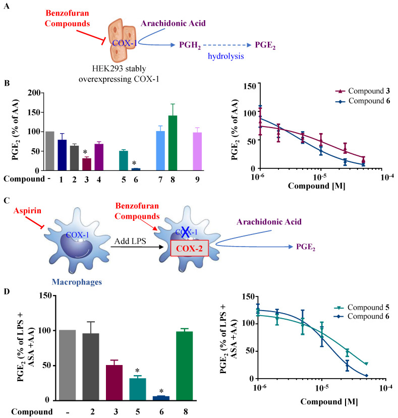
Figure 3.
Effects of the fluorinated benzofuran and dihydrobenzofuran on COX-1 and COX-2 activities. (A) Illustration for COX-1 activity assay. HEK-293 cells stably overexpressing COX-1 are treated with the nine derivatives of benzofuran at concentrations of only 10 µM for compounds 1, 7, and 9, and 50 µM for the other compounds, 30 min prior to the addition of 10 µM arachidonic acid (AA). This results in PGE2 production from PGH2 after hydrolysis, which reflects the COX-1 activity. (B) COX-1 activity. PGE2 formation was measured by enzyme immunoassay with the corresponding IC50 fitting curves for compounds 3 and 6. Levels of PGE2 were very low in the absence of arachidonic acid (0.04 ± 0.01 ng/mL) and 104.3 ± 16.7 ng/mL for 10 µM AA. (C) Illustration for COX-2 activity determination. Macrophages are treated with 10 µM of aspirin (ASA) for 30 min to block basal COX activity, washed, and treated with 10 ng/mL LPS for 24 h to induce COX-2. Cells are then washed and incubated 30 min with 50 µM of the different compounds prior to the addition of 10 µM of AA. The produced PGE2 mainly reflects COX-2 activity. (D) COX-2 activity. Percentage of LPS-treated cells was calculated, and data are represented as mean ± SEM (n = 4); * p < 0.05 versus AA for COX-1 activity, and versus LPS + ASA + AA for COX-2 activity (one-way ANOVA followed by Dunnett’s test). Corresponding IC50 fitting curves for compounds 5 and 6 are illustrated. Levels of PGE2 were very low in the absence of arachidonic acid (0.06 ± 0.01 ng/mL) and 71.7 ± 5.7 ng/mL for 10 µM AA.

