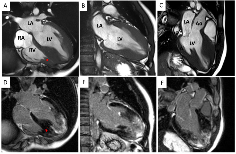
Figure 2.
Cardiac magnetic resonance morphological and fibrosis characterization. (A–C) Balanced Steady State Free Precession (bSSFP) cine 4−, 2− and 3−chamber views showing hypertrophy of the mid-apical segments of the left ventricle. Hypertrophy of the right ventricular wall and a right ventricular papillary muscle (red asterisk; panel (A)) is also noted. (D–F) Magnitude reconstruction (MAG) LGE 4−, 2− and 3−chamber views showing extensive late gadolinium enhancement (LGE) of the left ventricular mid-apical segments and the right (red arrow; panel (D)) ventricle. Ao = aorta, LV = left ventricle, RV = right ventricle, LA = left atrium, and RA = right atrium.

