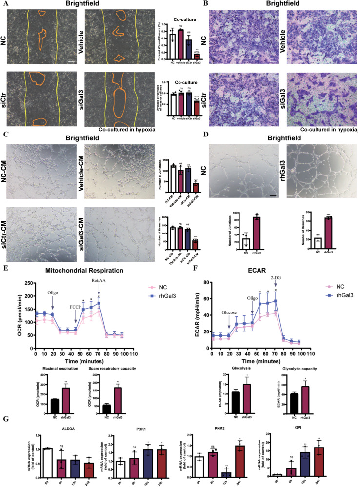
Fig. 4. Gal3 promoted endothelial proliferation and lifted endothelial metabolism.
A The bEnd.3 cells and were wounded with a p20 pipette tip and then were cocultured with BV2 transfected with siGal3 as compared to those with BV2 transfected with siRNA control and vehicle (lipo2000). Photographs were taken immediately and after 8 h (n = 3 per group for wound assay and two fields were calculated per sample). Scale bar, 100 µm. B The capacity of cell migration of bEnd.3 cells cocultured with microglia after 12 h in hypoxia was evaluated by transwell assay (n = 4 per group). Scale bar, 100 µm. C Lumen formation of bEnd.3 cells cultured by conditioned media was detected and compared between the groups after 6 h. (n = 3 per group). Scale bar, 100 µm. D The capacity of lumen formation of HUVECs cultured by normal ECM and ECM with rhGal3 after 6 h of stimulation (n = 3 per group). OCR (E) and ECAR (F) measured with the Seahorse XFe24 analyser in HUVECs cultured by normal ECM and ECM with Gal3 for 12 h (n = 5 per group). G mRNA level of enzymes in glycolysis was tested in HUVECs cultured by normal ECM and ECM with rhGal3 after 12 h by qRT-PCR, including ALDOA, PGK1, PKM2, and GPI. (n = 3 per group). Bars = means ± SD; *P < 0.05; **P < 0.01; ***P < 0.001, NS no significance.

