Fig. 1.
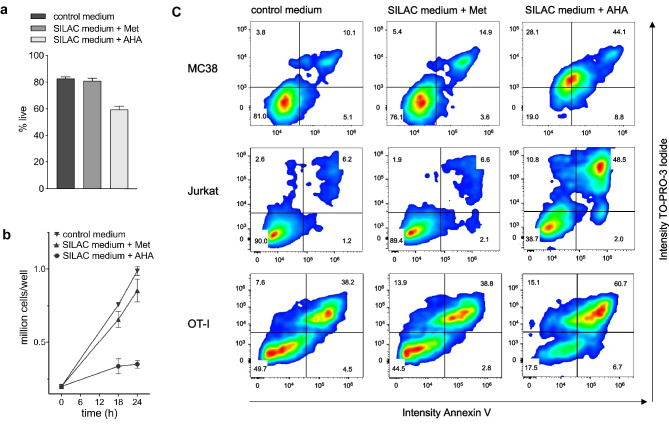
Influence of AHA on cell viability and growth. a MC38 cells were grown in control medium and medium containing Met or AHA for 20 h. Cell viability was determined by eFluor780 staining. b MC38 cells were cultured in control medium and medium containing Met or AHA. At the indicated time points, live cells were counted using trypan blue staining. c Indicated cells were cultured for 20 h in control medium and medium containing Met or AHA. The percentage of living and apoptotic/dead cells was determined by Annexin V and TO-PRO-3 Iodide staining followed by flow cytometry analysis

